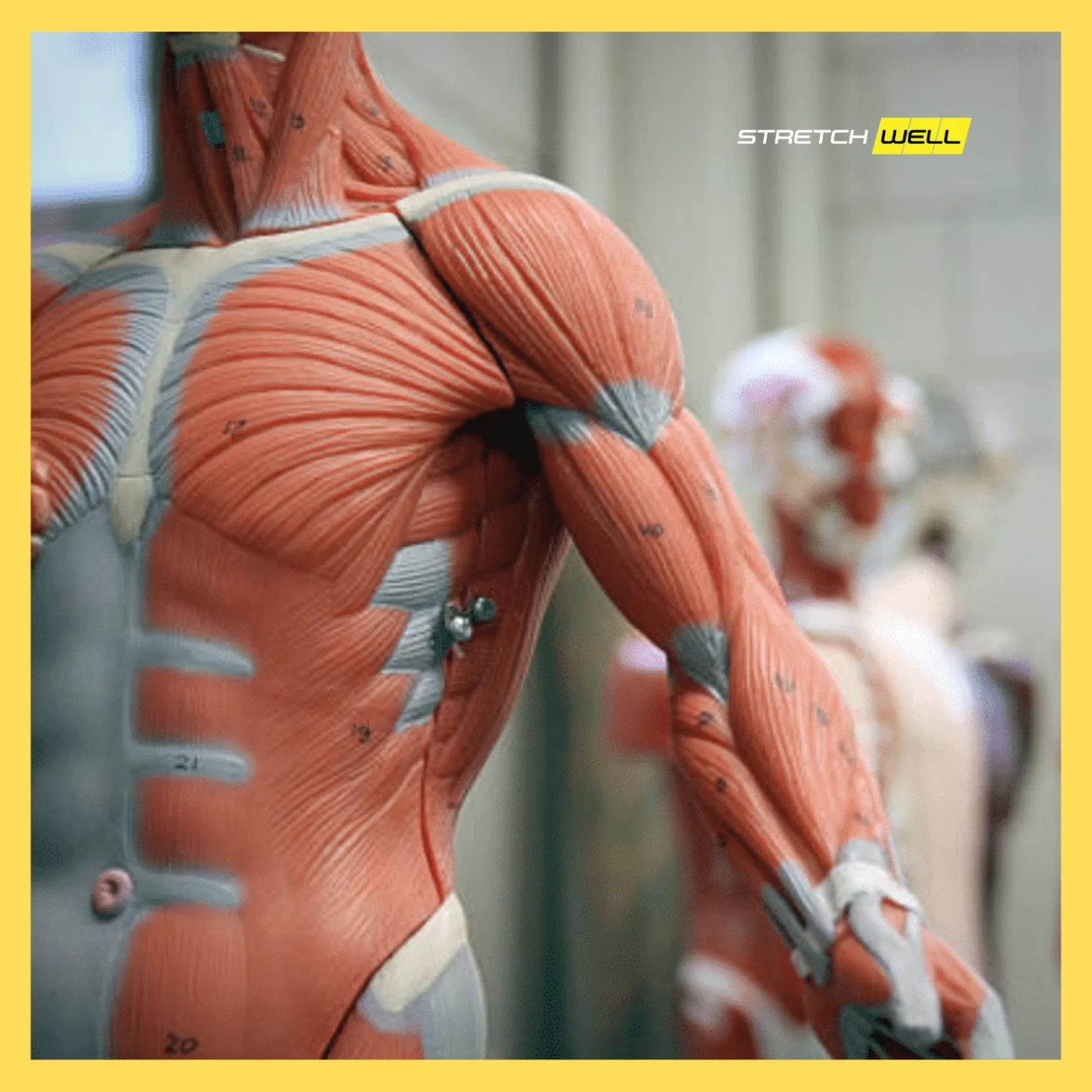
Muscle is a soft tissue, made up of special fibers (known as muscle fibers) that have the ability to contract. Muscle fibers are made up of a special type of cell called myocytes, which contain strands of actin and myosin proteins. As the threads cross each other, their muscle activity creates a contraction that changes the length and shape of the cell.
Muscle types:
– skeletal (striated),
– smooth muscles,
– heart muscle
Transverse-striated muscles

The striated (skeletal) muscles are attached to the bones and give shape to the body. Their cells are elongated in shape and without extensions, they have multiple nuclei that are located peripherally – under the cell membrane. They are among the largest cells in the body (up to 4 cm in humans) and make up about 40% of the total body weight. The membrane of these muscle cells is called the sarcolemma. The characteristic transverse-striation is reflected in alternating dark and light bands, which originates from the structure of the myofibrils themselves. Myofibrils are made up of thin actin and thick myosin fibers. The dark stripe is made up of actin and myosin, while the light stripe is made up of actin. In the middle of the light stripe there is a dark line called the Z-line. Skeletal muscles are innervated by the peripheral nervous system so that they work under the influence of our will and get tired during work. The primary function of these muscles is to move and produce body heat. Their muscle fibers are grouped into bundles that are wrapped in a connective membrane. Multiple bundles are combined into larger bundles, which are grouped and connected by a common connective membrane to build muscle.
Smooth muscles

Smooth (visceral) muscles build the walls of tubular organs and organs with cavities (intestines, stomach, bladder, blood vessels, uterus, etc.). They consist of a number of spindle-shaped cells that usually have a single nucleus in the center of the cell. Actin and myosin are grouped in bundles in them so that there is no transverse-striping. They are innervated by the vegetative (autonomic) nervous system so that they work outside the influence of our will. Innervation is dual, which means that each muscle is connected to both the sympathetic nervous system and the parasympathetic nervous system, which regulates the functioning of the organ (if the sympathetic nervous system accelerates the work of an organ, then the parasympathetic nervous system will act in the opposite way). Smooth muscle contractions are slow, last longer and there is no fatigue.
Heart muscle

The heart muscle is similar in structure to striated muscles, and in function it is more similar to smooth muscles. It is striated, but it is specially constructed as a physiological syncytium. The cells are abbreviated spindle-shaped and have extensions through which they connect to form a syncytium, which allows unhindered transmission of stimuli through the heart muscle (see figure). The contractions of the heart muscles are fast, rhythmic and automatic. There are two types of muscle cells in the heart:
• the single, typical muscle cells that make up the atria and ventricles of the heart, and
• others – Purkinje cells, which build the so-called automatic (conduction) system of the heart.
A syncytium is a large multinucleated cytoplasm that is delimited by a cell membrane but is not separated into individual mononuclear cells by inner membranes.
Muscle properties
1. Irritability (excitability) – The property of generating and implementing action potentials. This is due to the high peace potential of the membrane, which is -90 mV.
2. Contractility – Made possible by the presence of a contractile apparatus, which is made up of alternately placed and partially folded protein filaments of myosin and actin.
3. Stretchability – The ability of muscles to stretch to a greater length than that and at rest.
4. Elasticity – Muscles contain elastic elements that act to return to the initial length (serially and in parallel).